Imagine a routine kitty cold turning serious. Sometimes the common feline coronavirus (a common cat virus) mutates and becomes feline infectious peritonitis (FIP), a dangerous illness that damages tissues and organs. You might see a puffy, fluid-filled belly, or more subtle signs like quiet weight loss, fever, and low energy. Ever watched your cat suddenly stop jumping? I once watched Luna go from six-foot leaps to flopping on the couch, and that sudden change was a red flag. FIP usually hits young cats, about 6 to 24 months old, and it can also cause eye or brain problems that change how we treat things.
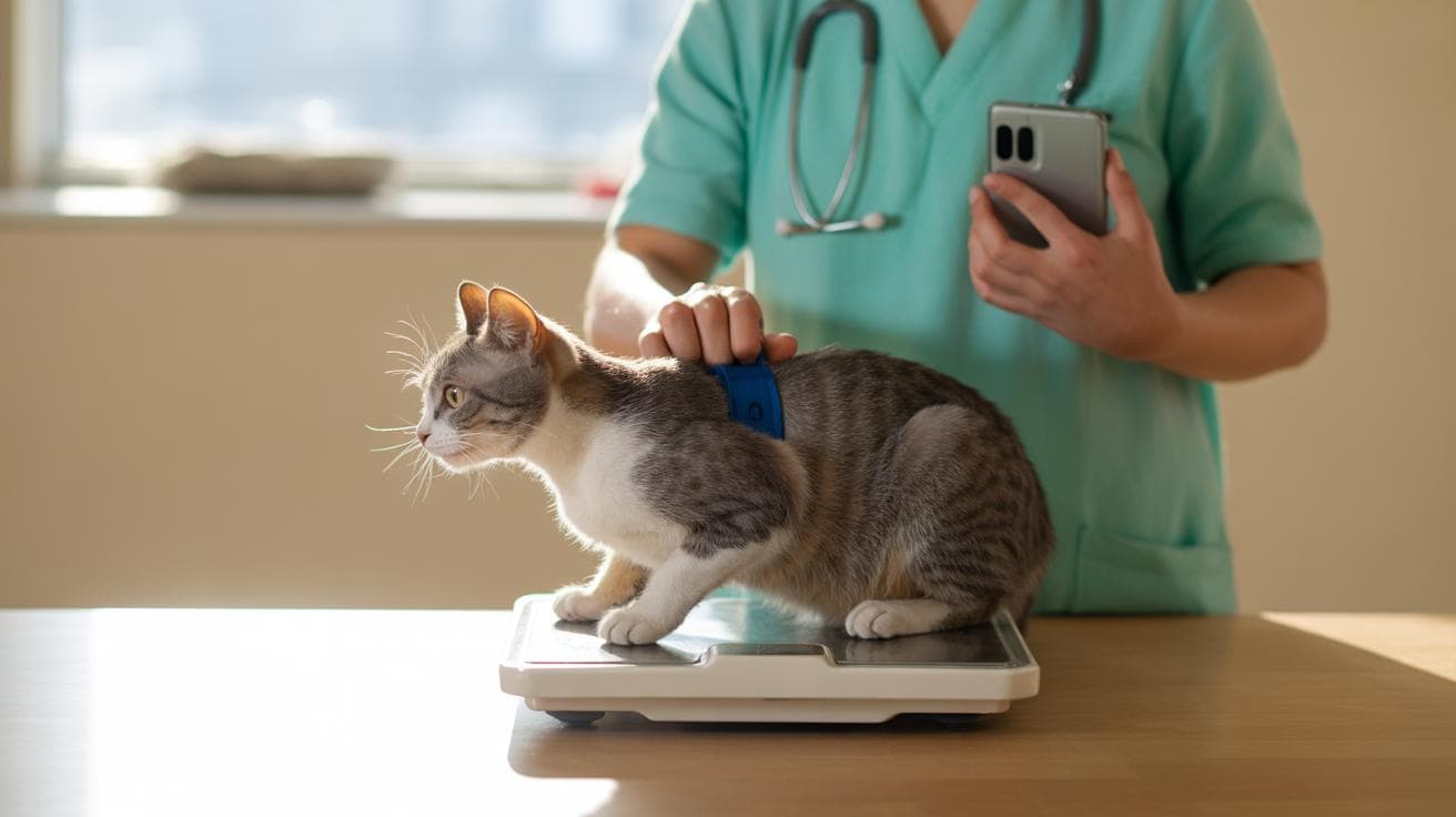
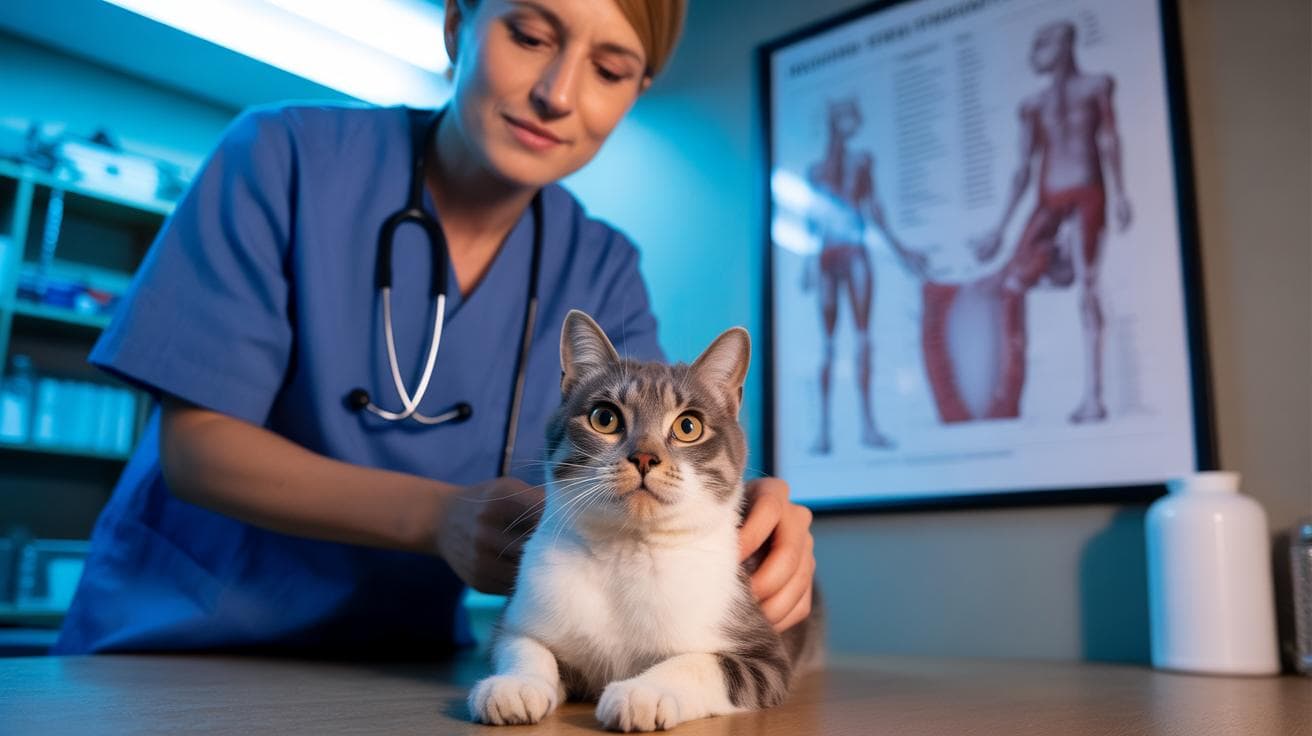
Diagnosing FIP can be tricky because its signs mimic other diseases. Your vet will use your cat’s history and a physical exam plus blood work (checks organ function and inflammation), ultrasound (sound-wave imaging) or X-rays, and sampling of any fluid for lab tests (cell counts, protein, virus checks). There are specific tests that look for the mutated virus or its effects, but often the diagnosis is a puzzle made from several clues. So expect follow-up visits and some detective work.
Treatment focuses on antiviral drugs (medications that stop viruses from multiplying) together with supportive care (fluids, nutrition, fever control, and pain relief). New antivirals have helped many cats recover, but treatment can be long and costly, and close monitoring is essential. Vets will tailor therapy to your cat’s symptoms and adjust if side effects appear. It’s not always simple, but many owners and vets have seen real improvements.
If FIP reaches the nervous system or the eyes you might notice stumbling, head tilts, seizures, circling, or vision changes, and those signs need urgent attention. Keep a close eye on behavior, appetite, and mobility, and call your vet if things change quickly. It’s scary, uh, but early diagnosis and treatment give the best chance for your kitty to feel feline fine again.
FIP in cats (feline infectious peritonitis): Diagnosis, Care
Feline infectious peritonitis, or FIP, is a serious illness caused when feline coronavirus (FCoV, the common cat coronavirus) mutates and starts damaging tissues. It usually shows up in two main ways: the wet form with fluid buildup, and the dry form with lumps or organ inflammation. Early veterinary care really matters, so read on for Diagnosing FIP, Treatment options, Monitoring/Prognosis, Causes/Pathophysiology, and Neurologic and ocular manifestations.
Key points:
- Two forms: wet (effusive, with fluid in the belly or chest) and dry (noneffusive, with inflammatory lesions in organs).
- Common signs: weight loss, fever, low energy; wet cases often have a swollen belly or trouble breathing.
- Most cats affected are young, usually 6 to 24 months old.
- Eye (ocular) and brain/nerve (neurologic) signs can happen and change how we diagnose and treat things.
- Antivirals (drugs that stop virus replication) can lead to remission for many cats.
- A definite diagnosis sometimes needs histopathology (microscopic exam of tissue samples).
- Prevention focuses on reducing FCoV spread in multi-cat homes or shelters.
- Relapse risk and best follow-up plans are still being studied.
Noticeable signs are often subtle at first. Your cat might seem quieter, eat less, or lose weight. Then you might spot a soft, round belly or faster breathing if fluid is building up. Ever watched your kitty suddenly stop jumping? Neurologic signs can look like stumbling, circling, or strange eye movements.
Treatment choices depend on the form and how sick the cat is. Antivirals, supportive care, and sometimes anti-inflammatory meds are the toolkit. Treatment options have improved a lot recently, but access and cost vary, so Practical Decisions matter , and, um, it’s okay to ask tough questions about affordability and goals of care.
Prevention helps in group settings. Good hygiene, minimizing overcrowding, and prompt testing of ill cats reduce FCoV spread. For shelters and multi-cat homes, simple steps can make a big difference.
Early vet checks are important. See your veterinarian right away if your cat has a swollen belly, breathing trouble, new neurologic signs, or unexplained weight loss. Check Diagnosing FIP for testing steps, Treatment options for therapies, and Practical Decisions for cost and access guidance.
Early action can change the story.
Causes and pathophysiology of FIP in cats
FIP starts when the common feline coronavirus, or FCoV, changes inside a cat and gains the ability to infect macrophages (immune cells that eat germs and move around the body). Those infected macrophages carry the virus into organs and trigger an over-the-top immune reaction that damages blood vessels – vasculitis (inflammation of vessel walls) – and leads to pyogranulomatous lesions (pus-filled, firm nodules). The funny, sad part is the damage comes more from the cat’s own immune response than from the virus itself, so symptoms depend on where the inflamed vessels and nodules show up.
FCoV spreads mostly by the fecal-oral route (cats pick up the virus from a contaminated litter box, food bowls, or hands), so the virus is very common in many cat groups. Studies find about 40% to 80% of cats have been exposed, and roughly one-third (around 33%) will shed the virus in feces at some point. Most cats either clear the intestinal infection or only shed briefly. A few shed for long periods, and only rarely does the virus mutate inside a cat and lead to FIP – that mutation can happen weeks, months, or even years after first exposure.
Young cats are the highest risk group. FIP usually appears between about 6 and 24 months of age, and estimates suggest around 5% to 10% of cats infected with FCoV go on to develop FIP, though overall FIP rates are often closer to 2% in many places. Crowded environments like catteries, shelters, or busy multi-cat homes, stressful changes, and some pedigree lines raise the odds. Intact males may have a slightly higher risk in some studies, so both environment and individual factors seem to matter.
Steps in how FIP develops
- FCoV infects the gut and replicates there.
- The cat may shed virus in feces for a short or long period.
- In a small number of cats the virus mutates and gains macrophage tropism (ability to infect macrophages).
- Infected macrophages spread systemically and cause immune-mediated vasculitis, which produces the clinical disease we call FIP.
Ever watched a kitten nap in the sun and thought, I hope it stays healthy? This is why understanding how FIP starts and spreads matters.
Clinical presentation of wet vs dry FIP in cats

FIP (feline infectious peritonitis) usually shows up in two main ways. One is the effusive or wet form, where fluid builds up in body cavities. The other is the non-effusive or dry form, which causes localized inflammation in organs. Lots of cats have signs of both. For CNS and eye-specific signs, see Neurologic and ocular manifestations.
Wet FIP signs:
- Abdominal swelling or ascites (ascites = fluid in the belly) that often feels squishy when you press the belly.
- A pot-bellied look with a tense, rounded abdomen.
- Trouble breathing from pleural effusion (pleural effusion = fluid around the lungs).
- Muffled or dull chest sounds when listening with a stethoscope (auscultation = listening to the chest).
- Fast clinical decline in severe cases , low energy and rapid breathing are common.
- The fluid is usually high in protein and straw-colored, and can be sticky or thick when analyzed.
Dry FIP signs:
- A persistent fever that doesn’t get better with ordinary antibiotics.
- Progressive weight loss and muscle wasting (loss of muscle mass) even if appetite hasn’t dropped much.
- Signs tied to specific organs, like jaundice (yellowing of skin and eyes) from liver involvement or renal azotemia (a rise in blood nitrogen waste, seen on blood tests) from kidney problems.
- Firm nodules or palpable masses in the abdomen caused by granulomatous inflammation (granulomatous inflammation = small, firm immune cell nodules).
- Eye problems such as uveitis (uveitis = inflammation inside the eye); see Neurologic and ocular manifestations for exam details.
- A course that can wax and wane, with signs that come and go over time.
| Feature | Wet FIP (effusive) | Dry FIP (non-effusive) |
|---|---|---|
| Typical fluid | Large-volume, high-protein ascites or pleural effusion | Minimal or no free fluid; focal organ lesions instead |
| Onset/progression | Often rapid decline over days to weeks | Slower, more variable progression over weeks to months |
| Most common clinical clues | Abdominal swelling, breathing trouble, muffled chest sounds | Fever, weight loss, organ-specific signs such as jaundice |
| Survival without treatment | Typically days to weeks in severe effusive cases | Often weeks to months, but variable by organ involvement |
Neurologic and ocular manifestations of FIP in cats
When feline infectious peritonitis hits the brain or the eyes, signs can come on fast and be pretty scary. These neurologic and eye changes often shift what vets choose to do and what the outlook looks like. If you think your kitty’s brain or eyes might be involved, check Clinical Presentation for overlapping systemic signs and follow the stepwise testing in Diagnosing FIP.
Neurologic signs are often obvious: seizures, wobbliness or ataxia (loss of balance), circling, a head tilt, weakness or partial paralysis (paresis), and cranial nerve problems that change pupils or swallowing. Advanced imaging like MRI (magnetic resonance imaging; detailed brain pictures) or CT (computed tomography; X-ray slice images) helps when there’s a focal lesion or suspected raised pressure. Analyzing cerebrospinal fluid (CSF = the clear fluid that bathes the brain and spinal cord) can show inflammation , more white blood cells and higher protein , although CSF isn’t always definitive. Referral to a neurologist and quick imaging can guide urgent care and safe sampling.
Eye problems usually show as anterior uveitis (inflammation inside the front of the eye), chorioretinitis (inflammation behind the retina), or sometimes retinal detachment (the retina pulling away, which can cause sudden blindness). An eye exam with fundoscopy (looking at the retina and back of the eye) often gives the key clues. Short-term treatment focuses on calming inflammation and protecting vision with topical eye medicines, systemic anti-inflammatory drugs when appropriate, and pain control. A timely referral to an ophthalmologist is a good idea. Antiviral choices should be discussed with your clinician in Treatment.
Warning signs of brain or eye involvement
- Seizures.
- Head tilt or other vestibular signs (losing balance).
- Sudden vision loss or bumping into things.
- Unequal pupils or odd pupil reactions (anisocoria).
- Light sensitivity or very small pupils suggesting anterior uveitis.
- A “dark curtain” across vision or abrupt blindness suggesting retinal detachment.
What vets often recommend
- MRI or CT for detailed brain imaging.
- CSF analysis to look for inflammatory cells and protein.
- Full eye exam with fundoscopy to check the retina.
- Acute treatments: anticonvulsants for seizures, anti-inflammatory therapy to calm the immune response, and topical ophthalmics for eye inflammation and comfort.
- Discuss antiviral options with your clinician as part of the overall plan.
If your cat suddenly seems off , like knocking into furniture or acting blind , don’t wait. These signs can be urgent. It’s tough to watch, I know, but quick diagnosis and the right referrals can make a real difference.
Diagnosing FIP in cats: tests, interpretation and diagnostic algorithm
FIP can feel like a mystery. No single noninvasive test proves it. So vets put together the history, physical exam, bloodwork, imaging, fluid or tissue analysis, PCR (polymerase chain reaction, a test that finds viral genetic material), and sometimes a biopsy to get a confident answer. This mix helps tell FIP apart from look-alike problems and lets you start the right treatment sooner.
Below is a practical roadmap you can use when you’re working up a sick cat. Think of it as a checklist to walk through, not a strict rulebook , and yes, I know it’s a lot. But step through these ideas and you’ll be clearer about next moves.
- Stabilize first. Fix dehydration, breathing trouble, or seizures, and run quick screening labs to spot life-threatening issues. Calm the cat, treat immediate problems, then dig into diagnostics.
- Get baseline bloodwork and imaging. Do a CBC (complete blood count) and chemistry panel, including the albumin-to-globulin (A:G) ratio (blood protein balance that often drops in FIP). Add an abdominal ultrasound and chest X-rays (thoracic radiographs) to look for fluid or organ changes.
- Collect and test any fluid or affected tissue. Tap effusions (fluid buildup) for cytology (cell-level exam), protein measurement, and FCoV PCR (tests for feline coronavirus RNA). Run a Rivalta test (a simple bedside drop test that hints at inflammatory fluid) if you have an effusion. Remember: positive PCR or Rivalta fits the story, but neither alone proves FIP because coronavirus and some infections can show the same results.
- When noninvasive tests are unclear, refer. Tissue biopsy and histopathology (microscopic tissue exam) with immunohistochemistry (IHC, a stain that shows viral antigen in cells) can confirm FIP. Referrals to internal medicine, neurology, or ophthalmology may be needed for tricky CNS or eye signs.
| Test | What it shows | Key limitations |
|---|---|---|
| CBC/chemistry & A:G ratio | Often sees high globulins (hyperglobulinemia), low A:G ratio (albumin to globulin), fewer lymphocytes (lymphopenia), and raised acute-phase proteins (inflammation markers) | Supportive but not specific. Other diseases can cause the same changes. |
| Effusion analysis & Rivalta | Effusive FIP usually gives thick, straw-colored, high-protein fluid. Rivalta (a quick drop test at the bedside) is often positive in inflammatory effusions. | Helpful for effusive cases but not definitive. Bacterial peritonitis and some other causes can look the same. |
| FCoV PCR (blood/effusion/tissue) | Detects viral RNA and can show a high viral load in effusion or tissue. | FCoV is common in cats. A positive PCR needs to be interpreted with the clinical picture to suggest FIP. |
| FCoV antibody testing | Shows prior exposure to feline coronavirus. | Many cats are antibody-positive, so this test by itself is not very useful for diagnosing FIP. |
| Imaging (X-ray/US, MRI for CNS) | Finds effusions, organ lesions, or brain/spinal changes if CNS is involved (MRI = magnetic resonance imaging, detailed soft-tissue pictures). | Suggestive findings but not proof. MRI is needed for focal CNS disease and may require referral. |
| Tissue biopsy / histopathology (IHC) | Gold standard: shows pyogranulomatous vasculitis (inflammatory blood vessel lesions) and viral antigen on IHC (a staining test that highlights virus in tissue). | Requires anesthesia and sampling or necropsy. Often needs referral and specialty lab work, but it’s the most definitive test. |
When should you refer? If noninvasive tests leave you unsure, if the cat has CNS or eye signs that need advanced imaging, or when a biopsy will change treatment choices, send the case on. Histopathology (microscopic tissue exam) looks for the typical pyogranulomatous vasculitis, and IHC (staining to detect viral antigen) is the confirmatory test. It can be done before death when safe, or at necropsy for final confirmation. Worth every careful step when the diagnosis matters.